
By Nicole Lasquete/AUGUST 3, 2026
In autoimmune diagnostics, laboratories routinely face a question that sounds simple but rarely is: which test should we run?
The answer is not one or the other. ELISA and indirect immunofluorescence assay (IFA) are complementary methods, each with distinct strengths, and each doing something the other cannot. Understanding where each belongs in the diagnostic pathway is what separates a workflow built for diagnostic confidence from one built on habit.
This guide explains how both methods work, what each does best, and how Immuno Concepts has designed them for the autoantibody testing ecosystem so that laboratories can make an informed decision.
What is ELISA?
ELISA, or enzyme-linked immunosorbent assay, is a plate-based immunological assay used to detect and quantify specific molecules in a biological sample, most commonly antibodies, antigens, proteins, or hormones.
It combines the specificity of antibody-antigen binding with the sensitivity of enzyme-amplified detection to produce a measurable, quantitative result.
How ELISA Works
The core process follows a consistent logic across all formats:
- Patient serum is added to microwells coated with a specific antigen
- If the target autoantibody is present, it binds to the antigen during incubation
- An enzyme-labeled secondary antibody then binds to the captured autoantibody
- A substrate solution triggers a color change whose intensity is directly proportional to the concentration of autoantibodies in the sample.
- Absorbance is measured on a microplate reader and converted to a calibrated antibody unit value.
Types of ELISA
ELISA is not a single format; it is a family of assays, each suited to different detection goals:
- Direct ELISA: The antigen is immobilized on the plate and detected by an enzyme-conjugated primary antibody. Fast and simple, but lower in sensitivity, and used primarily in research settings.
- Indirect ELISA: A primary antibody binds the antigen first; a labeled secondary antibody then amplifies the signal—the most widely used format in autoantibody testing and ANA screening.
- Sandwich ELISA: Two antibodies bind the target from opposite sides, delivering high specificity. Preferred for antigen detection, cytokines, growth factors, and infectious disease markers.
- Competitive ELISA: Labeled and unlabeled antigens compete for a fixed pool of antibodies. Useful for small molecules and haptens where sandwich formats are not applicable.
Applications of ELISA in Autoimmune Diagnostics
The ELISA immunoassay is used across a broad range of clinical and research applications:
- Confirmatory autoantibody detection following a positive ANA screen.
- Quantitative disease monitoring, tracking anti-dsDNA levels in SLE, anticardiolipin titers in antiphospholipid syndrome.
- ANCA specificity confirmation (MPO-ANCA and PR3-ANCA) following IFA screening.
- ENA profiling for Sjögren's syndrome, systemic sclerosis, and mixed connective tissue disease.
Immuno Concepts' RELISA® test systems apply the indirect ELISA format across a comprehensive range of autoantibody applications. Every RELISA® test kit ships with all components: antigen-coated microwells, conjugate, substrate, calibrator, controls, and buffers, validated together as one matched system. Every lot is tested against reference sera before release; if it does not meet Immuno Concepts' performance thresholds, it does not ship.
What is an Immunofluorescence Assay?
Immunofluorescence assay (IFA) is a laboratory technique that detects and visualizes specific antibodies or antigens in a patient sample using fluorescently labeled antibodies. Rather than producing a numerical value, IFA reveals a fluorescence pattern that carries direct diagnostic information about the type and distribution of autoantibodies present.
How IFA Works
- Patient serum is applied to a prepared substrate slide, typically HEp-2/2000® cells for ANA detection, or neutrophil substrates for ANCA testing.
- If autoantibodies are present in the serum, they bind to their corresponding antigens on the substrate during incubation.
- A fluorescently labeled secondary antibody is then applied to bind to any captured patient autoantibodies.
- The slide is examined under a fluorescence microscope; bound antibodies fluoresce, producing a pattern that is visually interpreted.
Some Applications of IFA in Autoimmune Diagnostics
- First-line ANA screening for suspected systemic rheumatic disease
- ANCA screening for ANCA-associated vasculitis using ethanol and formalin-fixed neutrophil substrates
- Anti-dsDNA detection using Crithidia luciliae substrate (the reference method for anti-DNA confirmation in SLE)
- Anti-endomysial antibody (EmA) detection for celiac disease
- Tissue-based autoantibody detection, ASMA, AMA, APCA, using rodent liver-kidney-stomach (LKS) substrates
Immuno Concepts has built its immunofluorescence assay portfolio around proprietary substrate technology developed over four decades. The HEp-2000® cell line: a patented substrate designed to dramatically increase sensitivity to SS-A/Ro autoantibodies without affecting other ANA patterns, delivers a clinically meaningful advantage for Sjögren's syndrome and other SS-A/Ro-associated conditions where standard HEp-2 substrates frequently miss the signal. For laboratories running high volumes of IFA, the Image Navigator® automated microscope from Immuno Concepts automates slide scanning, image capture, and pattern sorting, standardizing the most operator-dependent step in the workflow.
Similarities Between ELISA and Immunofluorescence Assay
Despite their different outputs and mechanisms, ELISA and IFA share several foundational characteristics:
- Both rely on antibody-antigen specificity: The ability of an antibody to bind its corresponding antigen with high selectivity is the basis of both assays, whether the output is a color change or a fluorescence pattern.
- Both are highly sensitive immunological assays: When properly validated and executed, both methods can detect autoantibodies at clinically relevant concentrations, including low-level positivity in early or mild disease.
- Both require validated reagent systems: The quality of substrates, conjugates, and controls directly determines the reliability of results in both assay types. Lot-to-lot consistency is a shared performance requirement.
- Both IFA and ELISA can be used in the diagnosis and monitoring of autoimmune diseases: They are standard tools in the diagnostic workup of conditions such as SLE, Sjögren's syndrome, systemic sclerosis, antiphospholipid syndrome, and ANCA-associated vasculitis.
Differences Between ELISA and Immunofluorescence Assay
While ELISA and IFA share the same foundational goal, detecting autoantibodies in patient samples, they diverge significantly in how they work, what they produce, and what clinical questions they are best positioned to answer.
Principle
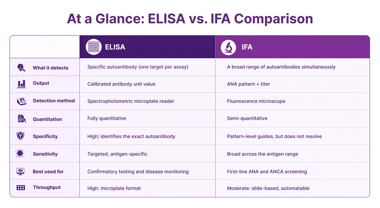
ELISA relies on the specific binding of a target autoantibody to an antigen-coated microwell, followed by enzymatic amplification of the captured signal. The enzyme catalyzes a color-producing reaction with a substrate, and the optical density of that color is measured and converted to a calibrated antibody unit value.
IFA relies on the specific binding of patient autoantibodies to antigens within a biological substrate, such as cells or tissue, followed by fluorescent labeling of the bound antibodies. The fluorescence pattern produced when the slide is examined under UV excitation carries the diagnostic information.
Detection Method
ELISA uses a spectrophotometric microplate reader to measure optical density. The result is an objective numerical output, a calibrated antibody unit value that can be compared directly across runs, lots, and time points. Immuno Concepts' RELISA® assays are specifically engineered to report these calibrated values, enabling results to be used for both initial diagnosis and longitudinal disease monitoring.
IFA uses a fluorescence microscope to detect and interpret the emitted fluorescent signal. The result is a pattern: homogeneous, speckled, nucleolar, centromere, cytoplasmic etc., which is read and classified by a trained operator. Immuno Concepts' Image Navigator® automated microscope reduces operator-dependent variability in this step by automating image capture, pre-sorting positives and negatives, and guiding pattern interpretation through Pattern Assisted Learning.
Output and Quantitation
ELISA produces a semi-quantitative result. Calibrated antibody unit values can be trended over time, making ELISA a popular choice for tracking disease activity over time and assessing treatment response. For example, rising anti-dsDNA values can signal a lupus flare, while declining values can indicate a good response to treatment through medication.
IFA produces a qualitative result: a titer and a pattern. The titer indicates the strength of the positive signal, while the pattern characterizes its clinical meaning. Both are diagnostic for disease association; the information relies on the reader's interpretation and is therefore subjective qualitative data. Despite this, a skilled reader can achieve similar performance to ELISA assays, including trending titers over time to predict changes in disease.
Specificity
ELISA delivers autoantibody-specific results. Each RELISA® test kit is designed around defined antigens, producing a result that identifies exactly which autoantibody is present. Two patients who share an identical ANA pattern on IFA may have entirely different autoantibody profiles; only ELISA confirmatory testing can distinguish them.
IFA provides pattern-level information. It broadly detects the presence and character of antinuclear antibody activity, but it does not determine which specific autoantibody is responsible for the observed pattern.
Sensitivity
ELISA is highly sensitive to its specific target antigen. The RELISA® ANA screen, for example, is calibrated with in-house-produced, well-characterized antigens for each target, delivering consistent, targeted sensitivity from lot to lot.
IFA simultaneously detects autoantibodies across a broad range of antigens, including those that may not be represented in a given ELISA panel. This breadth of detection is why IFA remains the recommended first-line method for ANA screening. Immuno Concepts' HEp-2000® substrate extends this sensitivity further for SS-A/Ro, an autoantibody that standard HEp-2 substrates frequently underdetect.
Throughput and Scalability
ELISA scales efficiently. Microplate formats enable the simultaneous processing of large numbers of patient samples. The RELISA® ENA Single-Well Screen coats each well with six ENA antigens simultaneously, enabling laboratories to exclude ENA-negative samples at high volume before committing to per-specificity confirmatory testing.
IFA is easily automated using Immuno Concepts' Image Navigator® automated microscope, and the Autoimmune FastTrack (AFT) IFA processor addresses throughput directly by automating liquid handling and image capture, improving both speed and consistency in high-volume IFA workflows.
Which Method Does Your Laboratory Need?
The right answer depends on what your laboratory is trying to achieve.
IFA delivers strong standalone value when:
- Broad, sensitive ANA screening is the priority, detecting the full picture of autoantibody activity from a single patient sample.
- Easily and efficiently rules out negative results for a broad spectrum of antibodies in the first screening test event.
- Pattern information is clinically important, guiding diagnosis based on whether the signal is homogeneous, speckled, nucleolar, centromeric, or cytoplasmic.
- ANCA screening or anti-DNA detection via Crithidia luciliae substrate is required
- The laboratory wants a visually rich, pattern-based result that communicates the character of autoantibody activity, not just its presence.
Immuno Concepts' immunofluorescence assay range, including HEp-2, HEp-2000®, ANCA, nDNA, and HISTOFLUOR® substrates, is built for exactly this. The HEp-2000® substrate in particular delivers significantly enhanced sensitivity to SS-A/Ro autoantibodies. This gives a clinically meaningful advantage for the detection of Systemic Lupus, Systemic Sclerosis, Sjögren's syndrome, and other rheumatic diseases via the detection of the SSA/Ro auto-antibody, which standard HEp-2 substrates routinely miss. The Image Navigator® automated microscope reduces operator dependence on pattern interpretation, making IFA scalable and consistent even at high volumes.
ELISA delivers strong standalone value when:
- Specific autoantibody identification is required, knowing not just that autoantibodies are present, but exactly which one.
- Quantitative output matters, calibrated antibody unit values that can be tracked serially to monitor disease activity or treatment response.
- High-throughput confirmatory testing is the workflow, processing large volumes efficiently without slide preparation and microscopy.
- The laboratory needs results that are directly comparable across runs, lots, and time points.
Immuno Concepts' RELISA® test systems are built for exactly this. In-house antigen production, pre-release lot validation, and matched reagent systems validated as one integrated unit ensure that RELISA® results are consistent, calibrated, and clinically dependable, whether the application is ENA profiling, anti-dsDNA quantification, anticardiolipin IgG and IgM determination, or ANCA specificity confirmation.
Take Your Pick Based on What Your Lab Needs
ELISA and immunofluorescence assay are both well-established immunological assays with real clinical value, and each can anchor a high-quality autoimmune diagnostic workflow on its own terms. IFA brings breadth, pattern intelligence, and visual diagnostic depth that no other method replicates. ELISA provides specificity, quantitation, and the longitudinal data that disease monitoring demands.
The right choice depends on what your laboratory needs to deliver. Immuno Concepts has spent over four decades building both immunofluorescence assays grounded in proprietary substrate technology and RELISA® test systems engineered for the consistency demanded by quantitative autoantibody testing. Whichever direction your laboratory is heading, Immuno Concepts has the products and the expertise to support it.