
By Nicole Lasquete/SEPTEMBER 24, 2026
Two patients can have the same ANA titer and share nothing else. What separates them is often not the number but the image: which cellular structures the antibodies bind to, and how that staining is distributed.
Immunofluorescence assays are widely used across diagnostic and research applications, enabling the visualization and localization of specific targets within cells and tissues. In clinical laboratories, their defining value is spatial: rather than reporting only whether an antibody is present, they show where the reaction occurs and what pattern it produces, information that can add important context to autoimmune testing.
That capability matters as positive results accumulate. Detection is only the beginning; the challenge is determining which findings warrant further investigation.
This article covers how immunofluorescence assays work, what they can and cannot establish, and the factors that determine the reliability of laboratory results.
What Is an Immunofluorescence Assay?
Immunofluorescence is a microscopy-based technique that uses antibodies and fluorescent labels to detect specific targets in cells, tissues, or other biological specimens. Clinical laboratories widely use immunofluorescence assays to visualize antibody reactions and support autoantibody testing.
Unlike methods that provide only a numerical or qualitative result, immunofluorescence assays preserve information about where the target is located within the specimen. In autoimmune diagnostics, this spatial information can add context for evaluating antibody reactions and determining whether further laboratory investigation is appropriate.
The exact approach depends on the specimen, target, substrate, and assay format, allowing immunofluorescence assays to support applications across clinical diagnostics, research, and other areas of laboratory science.
How Does Immunofluorescence Work?
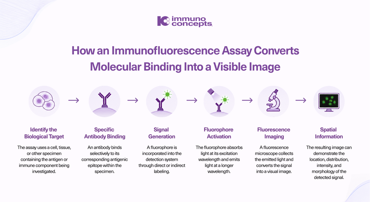
An immunofluorescence assay follows a sequence of steps that converts a specific antibody-target interaction into a detectable fluorescent signal. While the exact protocol varies by application, the underlying process remains consistent.
What Are the Different Types of Immunofluorescence?
Immunofluorescence assays can be configured in different ways depending on the target, specimen, and testing purpose. The primary formats are direct and indirect immunofluorescence.
Direct Immunofluorescence
Direct immunofluorescence (DIF) uses a fluorescently labeled antibody that binds directly to the target in a cell or tissue specimen. Because the detecting antibody already carries the fluorophore, the method involves fewer antibody-binding steps.
DIF is commonly used for tissue-based applications where the objective is to identify tissue-bound immunoglobulins, complement components, or other immune deposits in their original location.
Indirect Immunofluorescence
Indirect immunofluorescence (IIF) uses an unlabeled primary antibody followed by a fluorescently labeled secondary antibody. In autoimmune serology, the patient's antibodies can serve as the primary antibodies, binding to relevant targets on a prepared substrate.
Because multiple secondary antibodies can bind to a primary antibody, IIF can amplify the signal. It is widely used in autoantibody testing, including ANA testing with HEp-2 substrates.
The appropriate format depends on the target, specimen, assay objective, and detection requirements.
Why Does Substrate Choice Matter?
The substrate provides the biological environment where an immunofluorescence assay detects its target. It may consist of cultured cells, tissue sections, or other biological material selected to contain the structures relevant to the test.
Different substrates present different antigens and preserve cellular structures in different ways. As a result, substrate selection can influence both what an assay can detect and how the fluorescent reaction appears.
What Makes a Substrate Suitable?
- Target availability: The relevant antigen or structure must be present and accessible for antibody binding.
- Antigen preservation: Fixation and processing should preserve the target without significantly altering its accessibility.
- Cellular or tissue morphology: Well-preserved structures provide the context needed to interpret fluorescence.
- Signal-to-background: The substrate should clearly differentiate specific from nonspecific fluorescence.
- Reproducibility: Consistent substrate preparation helps reduce variation between assay runs.
In autoimmune diagnostics, substrate selection is particularly important. HEp-2 cells, for example, contain a broad range of nuclear and cytoplasmic structures and are widely used for ANA testing. Other applications may require specialized cells, tissue sections, or organisms that provide the appropriate targets.
The substrate is therefore an active part of the assay design, not simply a surface on which the reaction takes place.
What Can an Immunofluorescence Image Reveal?
The value of an immunofluorescence assay extends beyond detecting a fluorescent signal. The resulting image can show where a target is located and how it is distributed within the specimen.
What the laboratory observes depends on the target, substrate, specimen, and assay design. In autoantibody testing, for example, fluorescence may produce recognizable staining patterns that provide additional context for laboratory investigation.
Key Features of the Image
- Location: Where fluorescence appears within the cell or tissue.
- Distribution: How the signal is arranged across the specimen.
- Morphology: The shape and organization of the staining.
- Intensity: The relative strength of the fluorescent signal.
- Pattern: The overall configuration produced by the antibody-target interaction.
In autoimmune diagnostics, these characteristics can be particularly informative. ANA testing using HEp-2 cells, for example, can produce nuclear, cytoplasmic, and mitotic patterns classified using standardized nomenclature from the International Consensus on ANA Patterns (ICAP).
However, a staining pattern is not a diagnosis on its own. Its significance depends on the assay, specimen, antibody specificity, titer where applicable, and other clinical or laboratory findings.
What Factors Affect Immunofluorescence Results?
A clear fluorescent image does not automatically indicate a reliable result. Immunofluorescence assays depend on multiple variables across specimen preparation, antibody binding, staining, and image acquisition.
Antibody Specificity
The detection antibody must recognize the intended target with sufficient specificity. Excessive antibody concentration can increase nonspecific background, while insufficient concentration may produce a weak signal.
Fixation and Antigen Preservation
Fixation helps preserve cellular or tissue structures but can also affect antigen accessibility. The preparation method must therefore be appropriate for the target and assay design.
Background and Autofluorescence
Biological specimens may produce natural fluorescence, while nonspecific antibody binding can add unwanted signal. Excessive background can make specific staining more difficult to distinguish.
Imaging Conditions
Fluorophore selection, excitation intensity, exposure time, filters, and detector sensitivity can all influence the captured image. Prolonged exposure can also cause photobleaching and reduce signal intensity.
Controls and Standardization
Appropriate controls provide a reference for evaluating assay performance and distinguishing specific reactions from background. Consistent reagent handling, incubation, washing, and imaging conditions also help reduce variability.
Ultimately, result quality depends on the entire analytical workflow, rather than any single component of the immunofluorescence assay.
Where Are Immunofluorescence Assays Used?
The ability to visualize targets within their cellular or tissue context makes immunofluorescence assays useful across clinical diagnostics, biomedical research, and other laboratory applications. The specific use determines the specimen, substrate, detection format, and imaging approach.
Autoimmune Diagnostics
In autoimmune diagnostics, immunofluorescence detects and characterizes autoantibodies and immune deposits. Applications include ANA testing, ANCA testing, anti-dsDNA testing, and tissue-based investigations of autoimmune disease.
Tissue-Based Testing
Direct immunofluorescence can identify immunoglobulins, complement, and other immune components deposited within tissues. This can provide important information in investigations of autoimmune blistering diseases and certain renal conditions.
Research and Biomarker Studies
In research, immunofluorescence enables investigators to examine protein localization, cellular organization, signaling pathways, and other biological processes. Multiplex approaches can also visualize multiple targets within the same specimen.
The application ultimately determines what to detect, which substrate is appropriate, and how to evaluate the resulting fluorescence.
How Is Automation Changing Immunofluorescence?
As immunofluorescence assays move into higher-volume laboratory environments, automation can reduce repetitive manual work while supporting greater consistency across processing and imaging.
From Manual Steps to Connected Workflows
Automated systems can support tasks such as reagent dispensing, incubation, washing, slide handling, and fluorescence image acquisition. Digital imaging can also create searchable records for review, documentation, training, and quality assessment.
The goal is not to remove expert interpretation. Instead, lab automation allows professionals to spend less time on repetitive processing and more time evaluating complex or unusual findings.
For laboratories, the greater opportunity is workflow integration: connecting sample processing, imaging, review, and reporting rather than automating individual steps in isolation.
How Immuno Concepts Supports IFA Automation
For more than four decades, Immuno Concepts has focused on autoimmune diagnostics and technologies that support IFA workflows. Its portfolio includes next-gen lab automation solutions to support automated processing, digital fluorescence imaging, and connected analysis.
These technologies help laboratories reduce manual intervention, improve workflow consistency, and manage increasing testing demands while keeping laboratory expertise central to result review.