

By Nicole Lasquete/OCTOBER 9, 2026
Lupus can start with seemingly unrelated symptoms: persistent fatigue, joint pain, skin changes, unexplained fever, or symptoms that appear and disappear over time. This wide range of presentations is one reason lupus diagnosis can be challenging. According to the CDC, an estimated 204,000 people in the United States have systemic lupus erythematosus (SLE).
Lupus is a chronic autoimmune disease in which the immune system attacks the body's own tissues, causing inflammation that can affect multiple organs. Because its symptoms can overlap with other conditions, diagnosis relies on the complete clinical picture rather than a single symptom or lab result.
Autoantibody testing can provide important evidence during this evaluation. ANA testing, disease-associated antibody testing, blood counts, urine analysis, and other investigations may all contribute to the clinical assessment of suspected SLE.
This guide explores the major lupus types, common lupus erythematosus symptoms, and the laboratory testing used to evaluate suspected SLE, including ANA and anti-dsDNA antibody testing.
What Is Lupus Erythematosus?
Lupus erythematosus refers to a group of autoimmune conditions in which abnormal immune activity can cause inflammation and tissue damage. The most widely recognized form is systemic lupus erythematosus, which can affect the skin, joints, kidneys, lungs, heart, blood cells, and nervous system.
Unlike conditions that follow a relatively predictable course, lupus can vary considerably between individuals. Symptoms may become more pronounced during periods of disease activity and improve during periods of remission.
This variability is important when considering lupus diagnosis. A patient may initially present with nonspecific symptoms, while laboratory findings may provide additional clues that prompt more targeted investigation.
What Are the Different Types of Lupus?
Although lupus commonly refers to systemic lupus erythematosus, the term encompasses several forms with different clinical presentations.
Systemic Lupus Erythematosus
Systemic lupus erythematosus, or SLE, is the most common form of lupus. It can affect multiple organ systems, with symptoms commonly involving the skin and joints but potentially extending to the kidneys, lungs, heart, blood, and nervous system.
Since SLE can affect different organs in different patients, its presentation can vary considerably. This is one reason laboratory findings become an important part of the diagnostic evaluation.
Cutaneous Lupus
Cutaneous lupus primarily affects the skin. Patients may develop rashes or lesions, particularly in sun-exposed areas.
Some forms remain primarily skin-related, while cutaneous manifestations can also occur in people with systemic disease. Clinical examination and, when appropriate, laboratory or tissue-based investigations may help distinguish lupus-related skin findings from other conditions.
Drug-Induced Lupus
Drug-induced lupus is associated with exposure to certain medications. Its symptoms can resemble those of SLE but typically improve after the causative medication is discontinued under appropriate medical supervision.
A detailed medication history can therefore provide important context when evaluating lupus-like symptoms.
Neonatal Lupus
Neonatal lupus occurs when certain maternal antibodies cross the placenta and affect the developing fetus or newborn. It can be associated with skin, liver, or blood abnormalities and, in some cases, congenital heart block.
Unlike SLE, neonatal lupus is not caused by the infant developing an autoimmune disease in the same way. The condition is associated with maternal autoantibodies, so antibody testing and appropriate clinical monitoring are especially important.
Which Symptoms May Raise Suspicion for Lupus?
The wide range of lupus erythematosus symptoms can make the condition difficult to recognize. Symptoms may involve different parts of the body, vary in severity, and change over time.
Common Lupus Symptoms
Common lupus erythematosus symptoms can include:
- Fatigue
- Joint pain or swelling
- Fever
- Skin rashes
- Hair loss
- Mouth or nose sores
- Sensitivity to sunlight
Some patients may also experience changes in the fingers or toes associated with Raynaud's phenomenon.
These symptoms are not unique to lupus. Fatigue, joint pain, fever, and rashes can occur in many other conditions, so these symptoms alone do not establish a lupus diagnosis.
How Lupus Can Affect Different Organs
In SLE, inflammation can extend beyond the skin and joints.
Kidney involvement may produce protein or other abnormalities in urine. Lung or heart involvement may cause pain with breathing or other cardiopulmonary symptoms, while neurological involvement can produce a range of manifestations.
This broader range of possible findings explains why lupus evaluation often extends beyond symptom recognition. Laboratory testing can help identify immune markers and potential organ involvement that may not be apparent from symptoms alone.
Why Is Lupus Diagnosis So Complex?
There is no single test that independently confirms lupus. Instead, clinicians consider symptoms, medical history, physical examination findings, and laboratory results together.
A patient may initially present with nonspecific symptoms, such as fatigue or joint pain. When the clinical picture raises suspicion for SLE, ANA testing may provide an important starting point. Additional antibody testing and investigations for organ involvement can then add further context.
This stepwise approach is why lupus diagnosis depends on connecting clinical findings with multiple sources of laboratory evidence rather than relying on one result.
Which Blood Tests Support Lupus Diagnosis?
Laboratory testing can provide important evidence when SLE is suspected, but different tests address different aspects of the evaluation. Autoantibody testing can identify antibodies associated with autoimmune disease, while hematologic, complement, and urine testing can provide information about other findings relevant to the clinical assessment.
ANA Testing in the Evaluation of SLE
ANA testing is commonly performed when SLE or another systemic autoimmune disease is suspected. It detects antinuclear antibodies that react with components of the cell nucleus. ANA testing is highly sensitive for SLE, but a positive result does not establish the disease on its own. ANA positivity can also occur in people without lupus and in individuals with other autoimmune or medical conditions.
Immuno Concepts offers HEp-2 and HEp-2000® fluorescent test systems for ANA testing. Its HEp-2000® substrate is designed to increase sensitivity to SS-A/Ro autoantibodies while maintaining other ANA patterns.
For a deeper explanation of the methodology, see this guide to ANA testing.
Anti-dsDNA Testing
When ANA findings and the clinical presentation support further investigation, clinicians may evaluate anti-double-stranded DNA antibodies.
Anti-dsDNA antibodies are associated with SLE and can provide additional information when evaluating a patient with findings suggestive of the disease.
Indirect immunofluorescence using Crithidia luciliae is a recognized reference method for confirming anti-DNA positivity in suspected SLE.
Immuno Concepts' nDNA Fluorescent Test System uses Crithidia luciliae as the substrate for detecting anti-DNA antibodies. The assay is also FDA-cleared for use with the Image Navigator® system, allowing the testing workflow to connect with automated fluorescence imaging.
Additional Laboratory Testing
Other laboratory tests may provide additional information when evaluating suspected SLE. A complete blood count can identify abnormalities affecting red blood cells, white blood cells, or platelets. Complement testing may provide information about immune activity, while antiphospholipid antibody testing may be considered when clinically appropriate.
Immuno Concepts also offers RELISA® testing systems that can complement IFA-based testing. The RELISA® ANA Screen detects antibodies to multiple nuclear antigens, while RELISA® dsDNA provides a semi-quantitative method for detecting anti-dsDNA antibodies.
The choice of tests depends on the patient's presentation and the clinical questions being investigated.
How Does ANA Testing Contribute to Lupus Diagnosis?
ANA testing is often an early step in evaluating suspected SLE, but its value extends beyond a simple positive or negative result.
With indirect immunofluorescence, clinicians apply patient serum to a substrate containing a broad range of cellular antigens. If antinuclear antibodies are present, they bind to their corresponding targets. A fluorescently labeled secondary antibody then makes these interactions visible under a fluorescence microscope.
The resulting fluorescence can be evaluated for intensity and pattern, providing additional information that may help guide further testing.
Understanding ANA Results
A positive ANA indicates that antinuclear antibodies have been detected above the laboratory's reporting threshold. These antibodies can be strongly correlated with autoimmune disease; however, a positive result does not guarantee the individual has the disease. In a patient with symptoms compatible with lupus and a positive ANA result, this raises the overall suspicion for autoimmune disease, which would prompt further investigation and/or testing.
A negative ANA makes SLE less likely, given the high sensitivity of ANA testing, although results should still be interpreted within the broader clinical context.
Neither result should be considered diagnostic on its own.
Why ANA Patterns Matter
IFA can reveal fluorescence patterns that provide additional information about the autoantibodies present. Patterns such as homogeneous or speckled staining may be associated with different autoimmune conditions, although patterns alone cannot establish a specific diagnosis.
This is a key distinction between indirect immunofluorescence and methods that provide only an antigen-specific positive or negative result. IFA can provide visual information that helps guide subsequent laboratory investigation.
How Can Automated Microscopy Support IFA Testing for SLE?
For laboratories performing IFA as part of the evaluation of suspected SLE, consistent fluorescence image acquisition and review are important parts of the testing workflow. As testing volumes increase, manually reviewing and documenting fluorescence images can also add time to the process.
Image Navigator® is an automated microscope designed to capture, sort, and display fluorescence images from IFA slides. It can automatically acquire multiple high-resolution images, classify images into possible positive and possible negative categories, and provide a digital environment for review.
The system also supports barcode traceability, pattern libraries, ICAP integration, titer review, and connectivity with laboratory information systems. These capabilities can help laboratories organize the IFA image-review process while keeping laboratory professionals involved in pattern interpretation and final result confirmation.
Bringing Greater Clarity to Lupus Diagnosis
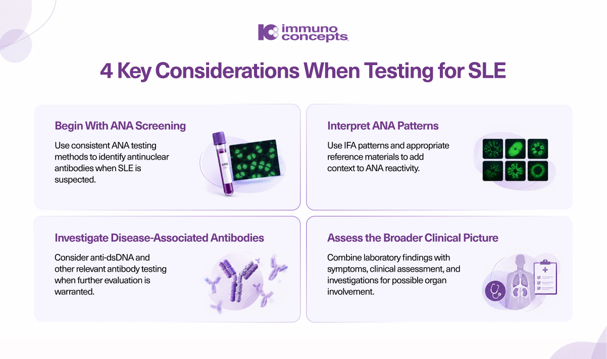
Lupus rarely follows a single diagnostic path. Symptoms may suggest an autoimmune condition, ANA testing can provide an initial signal, and additional antibody, blood, urine, and organ-specific investigations can help build a more complete picture.
That makes lupus diagnosis a process of connecting clinical findings with reliable laboratory evidence rather than relying on one definitive test.
For laboratories, supporting this process means having reliable methods for ANA screening, follow-up antibody testing, fluorescence pattern evaluation, and image review. Immuno Concepts provides IFA, ELISA, and automated microscopy solutions that can be incorporated into these different stages of autoimmune testing.
Together, these approaches can help laboratories build testing workflows that support consistent analysis while keeping clinical interpretation at the center of the diagnostic process.