

By Nicole Lasquete/AUGUST 26, 2026
Autoimmune disease diagnosis depends on one thing above all else: reliable detection of autoantibodies. Among the many testing methods available to clinical laboratories, indirect immunofluorescence (IFA) continues to hold its place as the reference standard. According to research, indirect immunofluorescence on HEp-2 cells is recognized by the American College of Rheumatology (ACR) and the International Consensus on ANA Patterns (ICAP) as the gold standard for antinuclear antibody screening. Despite the rise of newer immunoassay technologies, laboratories worldwide continue to rely on indirect immunofluorescence because alternative methods have shown inconsistent sensitivity and diagnostic accuracy.
This guide explains what indirect immunofluorescence is, how it works, and why it remains the preferred method for autoantibody testing in autoimmune diagnostics.
It also explores how Immuno Concepts has supported laboratories worldwide with high-quality IFA solutions that deliver consistent, reproducible results across a wide range of autoimmune diagnostic applications.
What Is Indirect Immunofluorescence?
Indirect immunofluorescence, often abbreviated as IFA or IIF, is a laboratory technique used to detect and characterize autoantibodies in a patient's serum. Unlike direct immunofluorescence, which uses a single labeled antibody, an indirect immunofluorescence test uses a two-step process: an unlabeled primary antibody (from the patient's serum) binds to a substrate antigen, and a fluorescently labeled secondary antibody then binds to that primary antibody. This indirect, two-antibody approach is what gives the method its name and its diagnostic power.
Because indirect immunofluorescence can detect a broad range of autoantibodies on a single substrate, identify the specific staining pattern, and determine an antibody's titer, it provides a depth of diagnostic information that few other methods can match.
How Does an Indirect Immunofluorescence Assay Work?
An indirect immunofluorescence assay follows a carefully controlled workflow that enables laboratories to detect and visualize disease-specific autoantibodies with exceptional sensitivity. While the process is straightforward, every step contributes to producing reliable, clinically meaningful results.
Step 1: Antibody Binding
The patient's serum is applied to a slide containing a prepared biological substrate, such as HEp-2 cells, HEp-2000®, Crithidia luciliae, neutrophils, or tissue sections. If autoantibodies are present, they bind to their corresponding antigens on the substrate during incubation.
Step 2: Fluorescent Labeling
After excess antibodies are removed through washing, a fluorescently labeled secondary antibody is applied. This secondary antibody binds specifically to any patient antibodies attached to the substrate. Because multiple secondary antibodies can bind to a single primary antibody, the fluorescent signal is amplified, improving the assay's sensitivity.
Step 3: Pattern Visualization
Following a final wash and mounting procedure, the slide is examined under a fluorescence microscope. Distinct fluorescence patterns, including nuclear, cytoplasmic, centromere, nucleolar, and mitotic staining, become visible. These patterns provide valuable diagnostic information that helps clinicians identify specific autoimmune diseases and determine appropriate follow-up testing.
Modern laboratories increasingly integrate automated slide processors and digital imaging systems into this workflow to improve standardization, reduce manual variability, and increase throughput while preserving the diagnostic strengths of indirect immunofluorescence.
Understanding Common Fluorescence Patterns
One of the greatest strengths of an indirect immunofluorescence assay is its ability to reveal distinct fluorescence patterns that provide valuable diagnostic insight. While a positive result confirms the presence of autoantibodies, the observed staining pattern can help narrow the differential diagnosis and guide additional confirmatory testing.
For example, when discussing the HEp-2/HEp-2000® assays, some of the most commonly recognized patterns include:
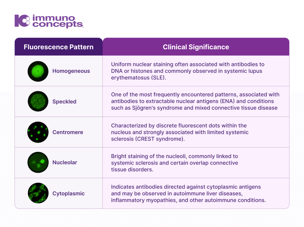
While fluorescence patterns alone are not diagnostic, they provide important clinical context that helps laboratory professionals and clinicians determine the most appropriate follow-up testing and interpretation.
Why Indirect Immunofluorescence Remains the Gold Standard
Although newer immunoassays have improved automation and throughput, indirect immunofluorescence remains recommended by leading professional organizations. Its ability to screen for a broad spectrum of autoantibodies while simultaneously revealing clinically meaningful fluorescence patterns sets it apart from many alternative testing methods. Rather than replacing antigen-specific assays, indirect immunofluorescence complements them by serving as a highly sensitive first-line screening method.
Broad Antigen Detection
An indirect immunofluorescence test using HEp-2-derived cells can detect antibodies against a wide array of nuclear and cytoplasmic antigens simultaneously, something single-antigen assays like ELISA cannot replicate on their own.
Pattern Recognition Adds Clinical Context
Beyond simply identifying the presence of autoantibodies, indirect immunofluorescence provides clinically meaningful fluorescence patterns that help narrow the differential diagnosis and guide confirmatory testing.
High Sensitivity
Studies evaluating automated and manual reading of ANA indirect immunofluorescence testing have reported sensitivity figures in the high 90s when compared against reference interpretation, reinforcing why regulatory and professional bodies continue to endorse it as the preferred screening method.
Titer Determination
Since indirect immunofluorescence uses serial dilution, laboratories can determine not just a positive or negative result but a quantitative titer, useful for monitoring disease activity over time.
Common Applications of Indirect Immunofluorescence Testing
Indirect immunofluorescence testing supports diagnosis across a wide range of autoimmune and connective tissue diseases. Some of the most common applications include:
- ANA (HEp-2/HEp-2000® substrates): Screening for antinuclear antibodies associated with systemic lupus erythematosus, Sjögren's syndrome, and other connective tissue diseases.
- Anti-dsDNA (Crithidia luciliae substrate): Confirming anti-DNA positivity in patients with suspected systemic lupus erythematosus.
- ANCA Testing (ethanol- and formalin-fixed neutrophil substrates): Screening for antineutrophil cytoplasmic antibodies associated with vasculitis, differentiating c-ANCA from p-ANCA patterns.
- Tissue-based autoantibody testing: Detection of smooth muscle antibodies (ASMA), mitochondrial antibodies (AMA), and parietal cell antibodies (APCA) using rodent tissue substrates.
- Anti-endomysial antibody (EmA) testing: Aiding in the diagnosis of celiac disease and dermatitis herpetiformis.
Indirect Immunofluorescence vs. Other Autoantibody Testing Methods
Laboratories frequently compare an indirect immunofluorescence test against solid-phase assays such as ELISA or multiplex bead-based platforms. While these alternative methods can offer faster processing and easier automation, published evaluations have found that they can vary meaningfully in sensitivity and diagnostic accuracy compared to HEp-2-based indirect immunofluorescence. This is largely due to differences in antigen source, purity, and the limited antigen panels used. This is a central reason professional bodies continue to recommend indirect immunofluorescence as the confirmatory and screening method of choice, with solid-phase assays often used as complementary rather than replacement tools.
Challenges in Indirect Immunofluorescence Testing
Despite its diagnostic strength, indirect immunofluorescence testing does come with operational challenges that laboratories must manage:
- Labor intensity: Manual slide preparation, incubation, and microscopic reading require skilled technologists and consistent technique.
- Inter-observer variability: Because pattern interpretation traditionally relies on human reading, results can vary between technologists and laboratories.
- Turnaround time: Manual indirect immunofluorescence workflows can be slower than automated solid-phase assays, particularly at high sample volumes.
This is where automation is reshaping the field. Automated slide processors and imaging systems now standardize incubation times, capture consistent digital images, and support more objective pattern classification, helping laboratories preserve the diagnostic strength of indirect immunofluorescence while improving throughput and reproducibility.
Choosing the Right Indirect Immunofluorescence Test System
When evaluating an indirect immunofluorescence assay or IFA test kit for your laboratory, consider the following:
- Substrate quality: Consistent, well-characterized substrates (such as HEp-2 or HEp-2000® cell lines) directly affect fluorescence clarity and pattern definition.
- Reagent integration: Buffers, conjugates, and mounting media validated together as a complete system reduce variability between runs.
- Automation compatibility: IFA test systems designed to work with automated processors and imaging platforms help laboratories scale testing volume without sacrificing accuracy.
- Application range: A comprehensive indirect immunofluorescence portfolio, spanning ANA, ANCA, anti-dsDNA, and tissue-based substrates, allows a single validated method to support multiple areas of autoimmune diagnostics.
- Manufacturer support: Lot-to-lot validation, training resources, and technical support help laboratories get consistent performance from every kit.
How Immuno Concepts Supports Indirect Immunofluorescence Testing
As laboratories continue to balance increasing testing volumes with the need for consistent diagnostic accuracy, choosing the right indirect immunofluorescence partner becomes just as important as selecting the right testing methodology. For more than 40 years, Immuno Concepts has focused exclusively on developing high-quality indirect immunofluorescence solutions that support reliable autoimmune diagnostics worldwide.
- HEp-2000® ANA: A patented, genetically engineered HEp-2 cell line with heightened sensitivity to SS-A/Ro autoantibodies, addressing a pattern frequently missed in conditions like Sjögren's syndrome.
- HEp-2 ANA: The original HEp-2 blend, produced to the same quality standards that established Immuno Concepts as a trusted name in indirect immunofluorescence testing.
- nDNA (Crithidia luciliae): The most widely used indirect immunofluorescence assay for anti-DNA detection, serving as the reference method for confirming anti-DNA positivity in suspected SLE cases.
- ANCA Ethanol and ANCA Formalin substrates: Supporting the complete ANCA testing algorithm for vasculitis screening and pattern differentiation.
- HISTOFLUOR® Rodent LKS and EmA substrates: Tissue-based indirect immunofluorescence testing kits supporting ASMA, AMA, APCA, and anti-endomysial antibody detection.
Every substrate is produced in-house using proprietary cell and tissue processes, and every lot is validated against reference sera before shipment. For laboratories building or scaling autoimmune diagnostic workflows, Immuno Concepts also offers automation-compatible solutions, including the DAS automated pipetting platform, the Image Navigator® automated microscope, and the Autoimmune FastTrack (AFT) IFA Processor, so laboratories can pair the diagnostic strength of indirect immunofluorescence with the consistency and throughput automation provides.
Building Diagnostic Confidence Through Indirect Immunofluorescence
As autoimmune diagnostics continue to evolve, indirect immunofluorescence remains the benchmark for sensitive, reliable autoantibody detection. Its ability to combine broad antibody screening with clinically meaningful fluorescence patterns makes it an indispensable tool for laboratories seeking accurate and consistent diagnostic results.
For laboratories seeking accurate and reproducible results, choosing high-quality IFA solutions remains essential to delivering confident clinical decisions.