

By Nicole Lasquete/NOVEMBER 18, 2026
Autoimmune diseases affecting the liver, bile ducts, and stomach can overlap, complicating diagnostic evaluation. In a study of 104 patients with autoimmune liver diseases, 22.1% tested positive for anti-parietal cell antibodies, and histological evaluation confirmed autoimmune gastritis in 91.3% of the APCA-positive group.
These findings illustrate why laboratory investigations may extend beyond the initially suspected organ system. Testing for anti-mitochondrial antibodies (AMA), anti-smooth muscle antibodies (ASMA), and anti-parietal cell antibodies (APCA) can provide important evidence when evaluating autoimmune liver and gastric conditions.
However, detecting these antibodies involves more than identifying a positive result. Their staining patterns, the tissue structures in which they appear, and the patient's broader clinical findings all contribute to interpretation. Rodent tissue-based immunofluorescence provides a method for examining these antibody reactions within relevant tissue structures.
This guide explores the clinical significance of AMA, ASMA, and APCA antibodies and the laboratory considerations that contribute to reliable autoimmune disease testing.
Why Are AMA, ASMA, and APCA Important in Autoimmune Disease Testing?
When a patient presents with unexplained liver abnormalities, persistent gastric symptoms, or findings suggestive of an autoimmune condition, clinicians may investigate whether the immune system is reacting against specific cellular structures. Autoantibody testing can narrow the diagnostic workup by identifying disease-specific immune responses.
AMA, ASMA, and anti-parietal cell antibodies (APCA) recognize different cellular targets and are associated with distinct autoimmune conditions. Indirect immunofluorescence on tissue substrates can help differentiate these antibodies by revealing characteristic staining patterns and their distribution across specific tissue structures.
Anti-Mitochondrial Antibodies (AMA)
Anti-mitochondrial antibodies react against antigens associated with mitochondria, the structures responsible for producing energy within cells. These antibodies are strongly associated with primary biliary cholangitis (PBC), a chronic autoimmune disease that damages the liver's small bile ducts.
An AMA blood test can help investigate suspected PBC, particularly when liver chemistry results or other clinical findings suggest cholestatic liver disease. However, AMA positivity alone does not establish the diagnosis. Clinical assessment and additional laboratory findings remain important, and some patients with PBC may be AMA-negative.
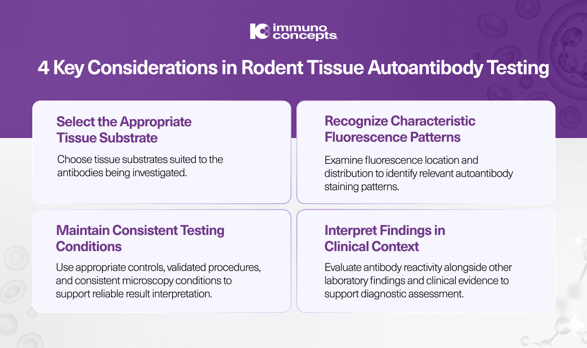
In rodent tissue, AMA may produce characteristic cytoplasmic fluorescence in liver cells and stain kidney tubular structures. Recognizing these patterns helps laboratory professionals distinguish mitochondrial reactivity from other antibody reactions.
Anti-Smooth Muscle Antibodies (ASMA)
Anti-smooth muscle antibodies target components of smooth muscle cells, particularly actin, although other cellular proteins may also contribute to the observed reactivity.
ASMA testing is associated with type 1 autoimmune hepatitis, a condition in which immune-mediated inflammation affects the liver. Clinicians may request an ASMA test when investigating unexplained elevations in liver enzymes or other findings suggestive of autoimmune hepatitis.
In rodent tissue, ASMA typically stains smooth muscle structures, including the muscular layers of the stomach and the walls of blood vessels in the liver. The appearance and distribution of staining help distinguish this reaction from mitochondrial or gastric parietal cell antibody patterns.
Still interpret ASMA findings alongside liver biochemistry, immunoglobulin levels, other autoantibodies, and relevant clinical or histological evidence.
Anti-Parietal Cell Antibodies (APCA)
Anti-parietal cell antibodies react against components of gastric parietal cells, including the hydrogen-potassium ATPase proton pump involved in gastric acid secretion. These antibodies are associated with autoimmune gastritis and may be detected when evaluating pernicious anemia, a condition involving vitamin B12 deficiency due to impaired absorption.
An anti-parietal cell antibody test can help identify an autoimmune response directed against the stomach lining. The result may prompt further evaluation of vitamin B12 status, iron levels, intrinsic factor antibodies, and other findings relevant to gastric function.
On rodent stomach tissue, APCA typically produces distinct fluorescence in gastric parietal cells. This localized staining pattern helps distinguish APCA reactivity from the broader tissue reactions associated with AMA and ASMA.
A positive APCA result does not independently confirm autoimmune gastritis or pernicious anemia. Some individuals have detectable antibodies without established clinical disease, making additional investigations necessary.
How Does Rodent Tissue Immunofluorescence Work?
Tissue-based antibody testing follows a sequence designed to reveal whether patient antibodies bind to particular structures in the tissue substrate.
In an indirect immunofluorescence assay, the lab incubates a patient serum sample with rodent tissue sections. If the relevant autoantibodies are present, they bind to their corresponding antigens in the substrate. After washing removes unbound antibodies, the lab adds a fluorescently labeled anti-human immunoglobulin reagent.
Under a fluorescence microscope, antibody-bound structures fluoresce. The location and distribution of this signal help the laboratory professional identify a pattern consistent with AMA, ASMA, or APCA reactivity.
For a closer look at the principles behind this methodology, explore our guide to immunofluorescence assays.
Recognizing the Staining Pattern
Fluorescence alone is not sufficient to establish a positive result. Laboratory professionals must evaluate whether the staining forms a discernible pattern consistent with the antibody being investigated.
For example, mitochondrial reactivity may appear across cytoplasmic structures and renal tubules, while APCA produces characteristic staining of gastric parietal cells. ASMA reactivity is evaluated by examining smooth muscle structures. The availability of these tissue structures within an assay depends on the substrate configuration.
The interpretation also considers background fluorescence and possible nonspecific staining. Comparing the patient result with appropriate controls helps determine whether the observed reaction matches the expected pattern.
Why Quality Controls Matter
Positive and negative controls help laboratories assess whether an assay is performing as expected. A suitable positive control should demonstrate the anticipated staining pattern, while a negative control helps identify nonspecific reactions. A buffer control can help assess nonspecific staining from the fluorescent reagent.
If controls do not meet the expected criteria, the run may be invalid and may require investigation or repetition, per the assay instructions.
Titratable controls can also help monitor run-to-run and lot-to-lot reproducibility. Consistent microscopy conditions, proper specimen handling, and adherence to the manufacturer's instructions further support dependable testing.
How Should AMA, ASMA, and APCA Results Be Interpreted?
A positive autoantibody result can support a diagnostic investigation, but it does not establish a disease independently. Interpretation depends on the antibody detected, the staining pattern, the clinical presentation, and findings from other investigations.
Interpreting AMA Results
AMA positivity can support the evaluation of primary biliary cholangitis, especially when liver chemistry findings are compatible with cholestatic disease. Additional assessment may include liver function testing, other disease-associated antibodies, imaging, or further investigations when indicated.
A negative AMA result does not completely exclude PBC. If clinical suspicion remains, clinicians may consider other relevant antibodies and diagnostic criteria.
Interpreting ASMA Results
ASMA positivity may support an investigation for type 1 autoimmune hepatitis, particularly when accompanied by elevated liver enzymes, increased immunoglobulin G, or other compatible findings.
ASMA can also occur in other clinical contexts. Its significance therefore depends on the complete diagnostic picture, and additional laboratory or histological evaluation may be necessary.
Interpreting APCA Results
APCA positivity can support an investigation for autoimmune gastritis and related conditions, including pernicious anemia. However, these antibodies may be detected in individuals without established disease and in people with other autoimmune conditions.
Depending on the clinical findings, further evaluation may include vitamin B12 and iron measurements, intrinsic factor antibody testing, gastrin measurements, or endoscopic and histological assessment.
Across all three tests, consider antibody findings as supporting evidence within a broader clinical evaluation. Laboratories report results according to the assay's validated interpretation criteria, while healthcare professionals determine their clinical significance.
How Can Laboratories Standardize Rodent Tissue Autoantibody Testing?
Reliable autoimmune disease testing depends on the quality of the tissue substrate, assay reagents, controls, microscopy conditions, and interpretation process. Differences in staining quality or inconsistent reading practices can complicate comparing results across runs.
Laboratories can support consistency by following validated procedures for specimen preparation, incubation, washing, and fluorescence microscopy. Appropriate positive and negative controls should be incorporated according to the manufacturer's instructions, and laboratory personnel should be trained to recognize expected staining patterns and potential sources of interference.
Documentation also matters. Recording the observed pattern, result, and relevant titer information, where applicable, supports traceability and reproducibility. Standardized microscopy settings are especially important when comparing fluorescence intensity or endpoint titers over time.
Supporting Image Review With Laboratory Technology
Manual fluorescence microscopy remains an established method for evaluating tissue-based autoantibody reactions. In laboratories managing higher testing volumes, digital image acquisition can also help organize the review process.
Immuno Concepts' HISTOFLUOR® Rodent LKS fluorescent test system is designed for the qualitative and semi-quantitative detection of IgG AMA, ASMA, and APCA in human serum. Its three-tissue configuration brings together rodent liver, kidney, and stomach substrates in one testing system, allowing laboratories to evaluate the characteristic staining patterns associated with all three antibody types.
The system works with the Image Navigator® automated microscope, which automates fluorescence image acquisition and sorting while allowing laboratory professionals to review and confirm results.
By combining tissue-based testing, appropriate controls, standardized microscopy, and trained interpretation, laboratories can support the evaluation of autoantibodies associated with autoimmune liver and gastric diseases.
Supporting Accurate Autoimmune Disease Testing
AMA, ASMA, and APCA provide different clues when investigating autoimmune conditions affecting the liver, bile ducts, and stomach. Their diagnostic value depends on recognizing the relevant antibody reaction and understanding how it fits into the patient's wider clinical presentation.
Rodent tissue-based immunofluorescence allows laboratories to examine characteristic autoantibody staining patterns within relevant tissue structures. Systems such as Immuno Concepts' HISTOFLUOR® Rodent LKS bring rodent liver, kidney, and stomach substrates together in one configuration, supporting the evaluation of AMA, ASMA, and APCA reactivity.
For laboratories evaluating these antibody patterns, the choice of assay and tissue substrate can influence how effectively the testing workflow supports observation and interpretation. Immuno Concepts provides tissue-based fluorescent test systems and laboratory imaging solutions designed to support these diagnostic investigations.